Laser Microscopy
Definition: a technique for generating microscopic images by scanning objects with a laser
More general term: microscopy
German: Lasermikroskopie
Categories:  vision, displays and imaging,
  methods
Author: Dr. Rüdiger Paschotta
Cite the article using its DOI: https://doi.org/10.61835/hwy
Get citation code: Endnote (RIS) BibTex plain textHTML
Laser microscopy is a class of techniques for generating microscopic images. In most cases, one uses laser scanning of some sample with a diffraction-limited laser beam. Scanning may be achieved by moving either the laser beam or the sample. Light carrying information on the sample may be generated in different ways, for example by scattering, through polarization changes, by second harmonic generation or by exciting fluorescence (→ fluorescence microscopy). The intensity of that light coming from the object is recorded for each point in the sample . From these data, images can be produced on a computer, and of course they can be stored in electronic form. Numerical methods can be applied to process the images, e.g. to enhance the contrast.

A frequently used imaging technique is based on a confocal geometry (→ confocal scanning microscopes), where the light from the focus in the sample is imaged (e.g. with a microscope objective) onto a pinhole, behind which the optical power is detected. This geometry suppresses the influence of light coming from other regions in the sample, e.g. from before or after the focus because such light can not efficiently pass through the pinhole. In effect, mainly the depth resolution is improved. The confocal principle is emphasized in the term confocal laser scanning microscopy, and discussed in more detail in the article on confocal scanning microscopes.
Instead of fluorescence, one may exploit acoustic effects of pulsed laser beams; the resulting method is called optoacoustic or photoacoustic microscopy [13].
Methods for Sub-diffraction Resolution
For most variants of laser microscopy, diffraction limits the possible beam waist radius of the scanning laser beam and thus the obtained image resolution. However, some techniques allow to beat the diffraction limit, and are sometimes called super-resolution microscopy or nanoscopy. One class of methods is based on non-uniform illumination in combination with a nonlinear photoresponse. Stimulated emission depletion microscopy (STED microscopy) (explained in the article on fluorescence microscopy, and related to the Nobel Prize in Chemistry 2014) is the most popular method of that type. Other techniques are based on certain fluorescent molecules, which preferably occupy certain parts of the specimen and can be localized very precisely (→ stochastic optical reconstruction microscopy, STORM).
Image Processing for Digital Microscopes
Images produced with later microscopes are usually not directly observed through an optical instrument, but on a high-resolution computer screen, for example. That makes it easy to let several people simultaneously view images.
The computer can then also be used for various auxiliary purposes:
- Instead of simply displaying recorded intensity values with a gray scale, display software can use false colors. Associated display parameters can be modified at any time when the raw data are kept.
- It is easy to precisely measure distances and sizes of objects on images displayed on a screen.
- Software-based image processing can in some respects increase the image quality, e.g. enhance image contrast and remove certain image distortions and artifacts caused by the optics.
- Software may identify differences between two images, for example taken before and after some manipulations on the sample.
- Digital images can easily be copied, stored and transmitted over large distances.
More to Learn
Encyclopedia articles:
Suppliers
The RP Photonics Buyer's Guide contains 15 suppliers for laser microscopes. Among them:


DRS Daylight Solutions
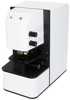
In a class of their own, the family of Spero® microscopes represents the world’s first and highest performing wide-field spectroscopic microscopy platform based on broadly tunable mid-infrared quantum cascade laser (QCL-IR) technology. Get high-quality data at unprecedented speeds.
Utilizing Daylight’s unique, patented QCL-IR light engine, Spero-QT 340 is faster than Raman and photothermal IR microscopes by orders of magnitude while avoiding sample auto-fluorescence and sample degradation by highly focused light sources.
Bibliography
| [1] | P. Davidovits and M. D. Egger, “Scanning laser microscope”, Nature 223 (5208), 831 (1969); https://doi.org/10.1038/223831a0 |
| [2] | P. Davidovits and M. D. Egger, “Scanning laser microscope for biological investigations”, Appl. Opt. 10 (7), 1615 (1971); https://doi.org/10.1364/AO.10.001615 |
| [3] | R. Kompfner et al., “Applications of Quantum Electronics; Part 1, the scanning optical microscope”, Oxford University Engineering Laboratory Report No. 1183177 (1975) |
| [4] | C. J. R. Sheppard and A. Choudhury, “Image formation in the scanning microscope”, Optica Acta 24 (10), 1051 (1977); https://doi.org/10.1080/713819421 |
| [5] | C. J. R. Sheppard and T. Wilson, “Depth of field in the scanning microscope”, Opt. Lett. 3 (3), 115 (1978); https://doi.org/10.1364/OL.3.000115 |
| [6] | J. N. Gannaway and C. J. R. Sheppard, “Second harmonic imaging in the scanning optical microscope”, Optical and Quantum Electon. 10 (5), 435 (1978); https://doi.org/10.1007/BF00620308 |
| [7] | I. J. Cox and C. J. R. Sheppard, “Scanning optical microscope incorporating a digital framestore and microcomputer ”, Appl. Opt. 22 (10), 1474 (1983); https://doi.org/10.1364/AO.22.001474 |
| [8] | W. Denk et al., “Two-photon laser scanning fluorescence microscopy”, Science 248, 73 (1990); https://doi.org/10.1126/science.2321027 |
| [9] | R. H. Webb, “Confocal optical microscopy”, Rep. Prog. Phys. 59 (3), 427 (1996); https://doi.org/10.1088/0034-4885/59/3/003 |
| [10] | J. Squier et al., “Third harmonic generation microscopy”, Opt. Express 3 (9), 315 (1998); https://doi.org/10.1364/OE.3.000315 |
| [11] | W. B. Amos and J. G. White, “How the confocal laser scanning microscope entered biological research”, Biology of the Cell 95 (6), 335 (2003); https://doi.org/10.1016/S0248-4900(03)00078-9 |
| [12] | J. A. Conchello and J. W. Lichtman, “Optical sectioning microscopy”, Nature Methods 2, 920 (2005); https://doi.org/10.1038/nmeth815 |
| [13] | L. V. Wang, “Multiscale photoacoustic microscopy and computed tomography”, Nature Photon. 3 (9), 503 (2009); https://doi.org/10.1038/nphoton.2009.157 |
| [14] | Nature Photon. 3 (7), special issue on super-resolution imaging (2009) |
| [15] | J. Bogusławski, A. Kwaśny, D. Stachowiak and G. Soboń, “Increasing brightness in multiphoton microscopy with a low-repetition-rate, wavelength-tunable femtosecond fiber laser”, Optics Continuum 3 (1), 22 (2024); https://doi.org/10.1364/OPTCON.505871 |
| [16] | C. J. R. Sheppard, “Confocal Microscopy. The Development of a Modern Microscopy”, Imaging & Microscopy (online, 2009) |
| [17] | National High Magnetic Field Laboratory, “Optical microscopy primer on fluorescence microscopy”, http://micro.magnet.fsu.edu/primer/techniques/fluorescence/fluorhome.html |
| [18] | K. W. Dunn et al., “Fundamental concepts in confocal microscopy”, http://www.microscopyu.com/articles/confocal/index.html |
| [19] | S. W. Hell, “Nobel Lecture: Nanoscopy with freely propagating light”, Rev. Mod. Phys. 87, 1169 (2015); https://doi.org/10.1103/RevModPhys.87.1169 |
Questions and Comments from Users
Here you can submit questions and comments. As far as they get accepted by the author, they will appear above this paragraph together with the author’s answer. The author will decide on acceptance based on certain criteria. Essentially, the issue must be of sufficiently broad interest.
Please do not enter personal data here; we would otherwise delete it soon. (See also our privacy declaration.) If you wish to receive personal feedback or consultancy from the author, please contact him, e.g. via e-mail.
By submitting the information, you give your consent to the potential publication of your inputs on our website according to our rules. (If you later retract your consent, we will delete those inputs.) As your inputs are first reviewed by the author, they may be published with some delay.


Connect and share this with your network:
Follow our specific LinkedIn pages for more insights and updates: